Research Article | | Peer-Reviewed

Molar-Incisor Hypomineralization (MIH) in an Ancient Population of Dor and Its Relation to the Aetiology of MIH

Received: 13 October 2024     Accepted: 30 October 2024     Published: 24 January 2025
Views:       Downloads:
Abstract

Background: The prevalence of MIH in modern population is increasing during the last decades. Most of the etiology factors today are based on perinatal health disorders. In order to understand if the hypomineralization of the enamel in MIH/HSPM is a novel developmental disorder, we compared the prevalence of modern population to archeological population with a high rate of mortality of young children. Objectives: To analyze the prevalence of MIH/HSPM in ancient population of Dor, Israel (16th-19th centuries). Materials: We examined 104 skulls out of 157 skeletons excavated, which had at least one permanent and/or one primary second molars for MIH/HSPM. Methods: All skulls were examined under a white light and skulls with MIH/SPMH were photographed. The skulls with suspected MIH/SPMH underwent CT analyses. Results: Three skulls out of the 104 skulls examined showed distinct enamel developmental defects on primary or permanent molars: one with MIH, one with HSPM and one with hypoplasia of second primary molars. Conclusions: Very low percentages of MIH/HSPM were found in Dor population, in comparison with modern prevalence of MIH in Israel, almost 18% of childrens 6-16 years old. Significance: In modern Israeli population with minimal perinatal health problems the prevalence of MIH/HSPM is very high and increasing. Based on the very low prevalence of MIH/HSPM in Dor population and the poor health situation of the children, we can conclude that the proposed aetiology of hypomineralization based on health or developmental problems during early childhood in modern population is questionable. Suggestion for further research: In modern populations the research should be directed to epigenetic factors in affected families.

Published in International Journal of Archaeology (Volume 13, Issue 1)
DOI 10.11648/j.ija.20251301.11
Page(s) 1-6
Creative Commons

This is an Open Access article, distributed under the terms of the Creative Commons Attribution 4.0 International License (http://creativecommons.org/licenses/by/4.0/), which permits unrestricted use, distribution and reproduction in any medium or format, provided the original work is properly cited.

Copyright

Copyright © The Author(s), 2025. Published by Science Publishing Group

Keywords

Molar-Incisor Hypomineralization (MIH), Ancient Populations, Hypoplasia, MIH Etiology

1. Introduction
Molar Incisor Hypomineralization (MIH) and Deciduous Molar Hypomineralization (DMH), also called Hypomineralization of Second Primary Molar (HSPM) describe a qualitative defect of the enamel due to hypomineralization of the ameloblasts during the perinatal period. The mineralization of second deciduous molars occurs from 18th weeks of pregnancy till 12 months after birth and during the first three years for the first permanent molars, but the clinical appearance of the hypomineralization of the deciduous or permanent molars can be observed after erruption, age 20-30 months for second deciduous molars and age 6-7 for first permanent molars . Becouse of that, the probable aetiology of hypomineralization was based on correlation of the clinical findings to health or developmental problems during early childhood, but all these reports are with low or very low quality of evidence . The proposed aetiology included perinatal hypoxia, prematurity and other perinatal problems, including caesarean section, and infant or childhood diseases of short duration. The only papers with high quality of evidence regarding aetiology of MIH are those dealing with epigenetic (monozygotic twins) and single nucleotid polymorphism analyses in patients and families . A systematic review analyzed the existing literature on genetic polymorphism associated with developmental defects of enamel . The conclusion was that MIH, HSPM dental fluorosis and hypoplasia have a complex etiology associated with genetic polymorphism of several genes. It is confirmed that the aetiology of MIH has a multifactorial aetiology in which duration, strength and timing of occurrence being responsible for the variable characteristics of the defect. Lately a paper by Garcia-Perez and associates correlated between MIH and body weigth among school-children. The prevalence of MIH ranges between 0.5-40% in different continents and countries . In South America is 18% and in Spain 21.1%. The prevalence of MIH among children 10 years of age or younger (15.1%) was much higher than the prevalence of MIH among older children (12.1%). The last modern pooled prevalence of the hypomineralization of permanent molars was 13.5% . America was the continent with highest prevalence (15.3%) and Asia had the lowest prevalence (10.7%). Affected incisors were observed in 36.6% of the cases. The prevalence of hypomineralization of the second deciduous molars was observed in 3.6% of the MIH cases. Modern populations shows a high prevalence of MIH, and the prevalence increased during the last decade by 2-11% in specific countries .
Regarding the increase in prevalence of MIH during the last decade, the question raised was: is MIH a novel developmental defect or can we find it in ancient populations, and at what prevalence? The prevalence of MIH in ancient population and the health status of the population can help us to understand which of the aetiology factors can be related today to MIH. Teeth have been a focus of interest for physical anthropologists over many generations. Teeth provide information about humans including cultural environment, locational migration, pathology, age estimation and sex differentiation . Pathologies, such as enamel developmental defects and caries are informative for understanding the health and nutrition status of individuals and populations . Several papers examined the prevalence of MIH in past populations. Curzon and associates described a medieval case of MIH from the 15th century . In a group of 323 skulls from Germany ( 12-20th century), 10 cases (3.1%) were classified as having MIH , 3.7% in 12-16th centuries, 9.1% in 16-18th centuries and 0% in 19-20th centuries. Garot and associates examined three skulls from France (7-18th centuries) with evidence of demarcated enamel hypomineralization on first permanent molars and primary molars.
The aim of this study was to evaluate the prevalence of MIH in an Ottoman period population from Palestine (16-19th centuries).
2. Materials and Methods
2.1. Dor-the Archeological Site
The location of Dor is 30 km south of the city of Haifa, on the Carmel coastal plain. Between the Middle Bronze Age and Roman periods, Dor was an important port. After the construction of the Caesarea port, Dor declined in importance. During the 3rd century, in the early Roman period, it was a fishing village. From the 4th to 7th centuries it served as an important Christian center with a church and associated structures. The Arab conquest during the 7th century caused the site to be abandoned. During the 16th through mid-19th centuries the deposits overlaying the Church were used for a cemetery. Archeological excavation carried out at the Byzantine church uncovered a number of skeletal remains buried in and around the church. They included individuals from the Byzantine and Ottoman periods, dated to the 16th to 19th centuries. Radiographs of the teeth were used to determine the age of the individuals using dental development for infant and children and dental attrition in adults . Age estimation using these procedures is accurate within a few months in the case of childrens, to within 2-3 years in adolescents, and within 10 years in adults. Only 157 individuals were sufficiently preserved for age estimation (Table 1). Out of these 157 individuals, only 104 squlls had at least one permanent or primary molar for analysis of hypomineralization.
Table 1. Age and sex distribution in the Dor population.

Gender/Age

0-1Y

2-5Y

7-10Y

11-17Y

18-24Y

25-39Y

40+Y

Unknown

Total

Female

2

7

4

14

13

40

Male

3

7

10

21

41

Unknown

30

13

10

2

1

2

16

74

Total No

30

13

15

16

5

26

34

16

157

%

19%

8%

10%

10%

3%

18%

22%

10%

100%

2.2. Dor-Health Status
The pathological lesions identified at Dor fall into two categories: developmental defects that results from acute infection or malnutrition during growth and development and/or signs of infectious diseases, trauma, tumors, degenerative diseases and nutritional disorders identified in the bones. At Dor enamel hypoplasia was present even in the primary teeth developing in utero. It indicate either chronic ill health and/or acute infection of a high percentage of pregnant woman. In permanent teeth enamel hypoplasia was present in 90% of all lower canines and 80% of upper canines, reinforcing the impression that growth stunning occurred in the Dor children. During the Otoman period malaria and tuberculosis were common and a major cause of death, followed by measels and enteric fevers. The paleopathology found at Dor indicates chronic anemia and poor calcium intake. Both the low life expectancy and the high prevalence of developmental insults inferred from the findings on enamel hypoplasia and growth arrest lines in long bones, indicate that health status at Dor was poor.
2.3. Clinical Examination for MIH/SPMH Findings at Dor
104 skulls from the excavation of DOR, which had at least one second primary molar and/or one permanent first molar, were examined clinically. All the teeth were examined and the findings classified according to the EAPD criteria for the diagnosis of MIH or HSPM. The examination was performed by a dentist on his 3rd year residency for specialization in pediatric dentistry with vast experience in diagnosis of MIH/SPMH. All cases were examined under a white light and skulls suspected to show MIH/SPMH were photographed and further analyzed by a specialist in pediatric dentistry with 40 years of clinical experience. The skulls with suspected MIH/SPMH underwent CT analyses.
3. Results
Three skulls out of the 104 skulls examined showed distinct enamel developmental defects on primary or permanent molars.
3.1. Case No 1
A 3-4 years old child. Enamel hypoplasia is present on the upper left primary central incisor, tooth #61 (Figure 1). Demarcated opacities can be observed on maxillary right second primary molar, tooth #55 and breakdown of enamel on the buccal surface of mandibular right second primary molar, tooth #85 (Figure 2). The CT slices shows the breakdown of enamel on teeth #55 and #85.
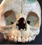
Figure 1. Frontal view case 1.
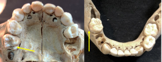
Figure 2. Occlusal views of the primary dentition.
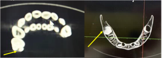
Figure 3. CT slices of the affected primary molars.
3.2. Case No 2
A 13-15 years old male with permanent dentition (Figure 4). Demarcated opacity with breakdown of enamel can be observed on the distal part of the upper left first molar, tooth #26 (Figure 5). No similar defects were observed on the other first molars. On the CT slice we can see the missing enamel on the palatinal surface of tooth #26 (Figure 6).
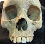
Figure 4. Frontal view case 2.
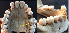
Figure 5. Occlusal view of the permanent dentition. See tooth #26.
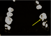
Figure 6. CT slice of tooth #26.
3.3. Case No 3
A 7-9 years old girl (Figure 7). A Tallon cusp can be seen on upper right lateral incisor, tooth #12. Primary molars with severe attrition, and hypoplasia on the occluso-palatal surface of second primary molars (Figure 8). On the CT image we can observe the missing enamel on the second primary molars (Figure 9).
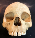
Figure 7. Frontal view case 3.
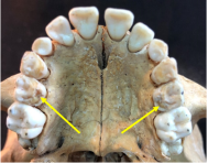
Figure 8. Occlusal view of the primary dentition. See the similarity of missing enamel on teeth #55 and #65 and the attrition of first primary molars.
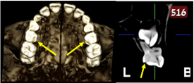
Figure 9. CT view of the occlusal surface of the dentition and slice of tooth #65.
4. Discussion
MIH and HSPM affects mineralization of primary and permanent molars and is sometimes associated with demarcated opacities on permanent incisors. The modern prevalence of MIH in Israel, the region of the archeological site of Dor, was almost 18% , and higher in the young age group compared to the older group. This tendency of higher prevalence in younger children was also observed in Mexico . In the archeological sites the diagnosis of hypomineralization is questionable. Taphonomic stains of teeth due to chemical elements contained in the burial ground can be similar to demarcated opacities of MIH . Another enamel defect that can mimic post eruptive enamel breakdown due to hypomineralization is enamel hypoplasia, a common enamel defect found in archeological sites . The differences between hypomineralization and hypoplasia of enamel are significant: hypoplasia is a quantitative defect of enamel that affects all the teeth developing during the same time period. The hypocalcification, a qualitative defect of the enamel, affects each molar tooth differently- from an intact tooth to a major breakdown of enamel in the same mouth. Surrounding the area of the breakdown of enamel, demarcated opacities can be observed in the same tooth. Garot and associates described three case of hypomineralization from archeological sites in France, two permanent molars affected by discoloration and reduced mineralization and one case of primary second molars with HSPM. Figure 2 in their paper shows four second primary molars affected by loss of enamel on the distal part of the crowns, disto-palatinal area in the upper primary molars and distal cusp in the lower primary molars, an area that develops at the same time during second primary molars development. So, by definition this case probably showed hypoplasia of enamel and not hypomineralization . The low prevalence of hypomineralization in archeological populations, 3.1% in Germany (16th-18th centuries) , or 1% of MIH and 1% HSPM in our research of the archeological population of Dor, compared to the high prevalence of MIH in modern populations, can indicate that the hypomineralization of the enamel increased during the last centuries. In the archeological site of Dor 47% of the individuals excavated were under the age of 10 years and the health status of the population was very poor . The death of the children from Dor was due to childhood diseases and malnutrition, and still only 1% of MIH and 1% of SPMH were present. So, all the modern perinatal aetiologies of MIH and HSPM described in Lygidakis and associates , like allergies, antibiotics, asthma, breast feeding, bronchitis, caesarean, celiac, chiken pox, diarrhoea, fever, fluoride, gastric disorders, gestational diabetes and hypertension, hypoxia at birth, kidney diseases, low birth weight, malnutrition, maternal diseases and more, should be looked at cautiously. More than that all these modern papers had low to very low quality of evidence .
5. Conclusions
The high prevalence of MIH in Modern Israelis compared to the low prevalence in Dor population shows that the prevalence is increasing lately, and based on the findings from Dor we can speculate that the aetiology of MIH follows genetic and/or epigenetic components. Further research shoud be aimed to genetic analysis of mutations in the genom of affected children in comparison with healthy family mmembers.
Abreviations

MIH

Molar Incisor Hypomineralization

HSPM

Hypomineralization of Primary Second Molar

Acknowledgments
To Prof. Patricia Smith from the Hebrew University, Jerusalem, for the opportunity to analyze the squlls from Dor excavation, and to Mr. Israel Dezuraev from Pano Bat-Yam for the CT analyses.
Author Contributions
Harel Sharon: Conceptualization, Methodology, Data curation, Investigation, Writing – original draft
Zilberman Uri: Conceptualization, Data curation, Methodology, Project administration, Supervision, Validation, Writing – review & editing
Conflicts of Interest
The authors declare no conflict of interest.
References
[1] Brook A. H., Smith J. M. 2006. Hypoplastic enamel defects and anvironmental stress in a homogenous Romano-British population. Eur. J. Oral. Sci. 114 Suppl 1: 370-374.
[2] Curzon M. E. J. et al. 2015. Case-report: A medieval case of molar-incisor-hypomineralization. Br. Dent. J. 219: 583-587.
[3] Damares Lego J. et al. 2022. MIH prevalence comparative study in 6 years of interval. Scientific World. J. 9: 4743252.
[4] Garcia Perez A. et al. 2024. Molar Incisor hypomineralization is associated with the prevalence of thinnes among schoolchildren in communities with different fluoride levels in the drinking water. Int. J. Dent. 1-8.
[5] Garot E. et al. 2017a. Analytical evidence of enamel hypomineralization on permanent and primary molars among past populations. Sci. Rep. 7: 1712.
[6] Garot E. et al. 2017b. Diagnostic guide enabling distinction between taphonomic stains and enamel hypomineralization in an archeological context. Arch. Oral. Biol. 74: 28-36.
[7] Goodman A. H. et al. 1980. Enamel hypoplasia as indicators of stress in three prehistoric populations from Illinois. Hum. Biol. 52: 515-528.
[8] Hassan J. et al. 2019. The prevalence of molar incisor hypomineralization among children in Jewish and Arab population in Israel. J. Israeli Dent. Associat. 36: 28-36.
[9] Irigoyen-Camacho M. E. et al. 2020. Evaluating the changes in molar incisor hypomineralization prevalence: A comparison of two cross-sectional studies in two elementary schools in Mexico City between 2008 and 2017. Clin. Exp. Dent. Res. 6: 82-89.
[10] Kuhnisch J. et al. 2016. Was molar incisor hypomineralizatio (MIH) present in archeological case series? Clin. Oral. Investig. 20: 2387-2393.
[11] Lopes L. B. et al. 2021. The prevalence of Molar-incisor hypomineralization: a systematic review and meta-analysis. Sci. Rep. 17: 11: 22405.
[12] Lopez-Fatturi A. et al. 2024. Genetic polymorphism associated with developmental defects of enamel: A systematic review. Int. J. Paediatr. Dent. 00: 1-13.
[13] Lygidakis N. A. et al. 2022. Best clinical practice guidance for clinicians dealing with children presenting with molar-incisor-hypomineralization (MIH): an updated European Academy of Paediatric Dentistry policy document. Eur. Arch. Paediatri. Dent. 23: 3-21.
[14] Miles A. E. W. 2001. The Miles method of assesing age from tooth wear revisited. J Archeological Sci. 28: 973-982.
[15] Moorrees C. F. A. et al. 1963. Age variation of formation stages for ten permanent teeth. J. Dent. Res. 42: 1490-1502.
[16] Smith P., Horwitz Kolska L. 2009. A synthetic approach to the study of diet, health and disease in an Ottoman period population from Palestine. In Al-Rafidan Journal of Western Asiatic Studies, volume XXX, 2009. The institute for cultural studies of ancient Iraq, Kokushien University, Tokyo.
[17] Stewart R. E. et al. 1982. General concepts of growth and development. In Stewart RE, Barber TK, Troutman KC, Wei SHY (eds). Pediatric dentistry: Scientific foundations and clinical practice. The CV Mosby Co. St Louis, Toronto, London. p. 19.
[18] Walter B. S. et al. 2015. Sex differentials in caries frequencies in Medieval London. Arch. Oral. Biol. 63: 32-39.
[19] Zhao D. et al. 2018. The prevalence of molar incisor hypomineralization: evidence from 70 studies. Int. J. Paediatr. Dent. 28: 170-179.
Cite This Article
  • APA Style

    Sharon, H., Uri, Z. (2025). Molar-Incisor Hypomineralization (MIH) in an Ancient Population of Dor and Its Relation to the Aetiology of MIH. International Journal of Archaeology, 13(1), 1-6. https://doi.org/10.11648/j.ija.20251301.11

    Copy | Download

    ACS Style

    Sharon, H.; Uri, Z. Molar-Incisor Hypomineralization (MIH) in an Ancient Population of Dor and Its Relation to the Aetiology of MIH. Int. J. Archaeol. 2025, 13(1), 1-6. doi: 10.11648/j.ija.20251301.11

    Copy | Download

    AMA Style

    Sharon H, Uri Z. Molar-Incisor Hypomineralization (MIH) in an Ancient Population of Dor and Its Relation to the Aetiology of MIH. Int J Archaeol. 2025;13(1):1-6. doi: 10.11648/j.ija.20251301.11

    Copy | Download

  • @article{10.11648/j.ija.20251301.11,
      author = {Harel Sharon and Zilberman Uri},
      title = {Molar-Incisor Hypomineralization (MIH) in an Ancient Population of Dor and Its Relation to the Aetiology of MIH},
      journal = {International Journal of Archaeology},
      volume = {13},
      number = {1},
      pages = {1-6},
      doi = {10.11648/j.ija.20251301.11},
      url = {https://doi.org/10.11648/j.ija.20251301.11},
      eprint = {https://article.sciencepublishinggroup.com/pdf/10.11648.j.ija.20251301.11},
      abstract = {Background: The prevalence of MIH in modern population is increasing during the last decades. Most of the etiology factors today are based on perinatal health disorders. In order to understand if the hypomineralization of the enamel in MIH/HSPM is a novel developmental disorder, we compared the prevalence of modern population to archeological population with a high rate of mortality of young children. Objectives: To analyze the prevalence of MIH/HSPM in ancient population of Dor, Israel (16th-19th centuries). Materials: We examined 104 skulls out of 157 skeletons excavated, which had at least one permanent and/or one primary second molars for MIH/HSPM. Methods: All skulls were examined under a white light and skulls with MIH/SPMH were photographed. The skulls with suspected MIH/SPMH underwent CT analyses. Results: Three skulls out of the 104 skulls examined showed distinct enamel developmental defects on primary or permanent molars: one with MIH, one with HSPM and one with hypoplasia of second primary molars. Conclusions: Very low percentages of MIH/HSPM were found in Dor population, in comparison with modern prevalence of MIH in Israel, almost 18% of childrens 6-16 years old. Significance: In modern Israeli population with minimal perinatal health problems the prevalence of MIH/HSPM is very high and increasing. Based on the very low prevalence of MIH/HSPM in Dor population and the poor health situation of the children, we can conclude that the proposed aetiology of hypomineralization based on health or developmental problems during early childhood in modern population is questionable. Suggestion for further research: In modern populations the research should be directed to epigenetic factors in affected families.},
     year = {2025}
    }
    

    Copy | Download

  • TY  - JOUR
    T1  - Molar-Incisor Hypomineralization (MIH) in an Ancient Population of Dor and Its Relation to the Aetiology of MIH
    AU  - Harel Sharon
    AU  - Zilberman Uri
    Y1  - 2025/01/24
    PY  - 2025
    N1  - https://doi.org/10.11648/j.ija.20251301.11
    DO  - 10.11648/j.ija.20251301.11
    T2  - International Journal of Archaeology
    JF  - International Journal of Archaeology
    JO  - International Journal of Archaeology
    SP  - 1
    EP  - 6
    PB  - Science Publishing Group
    SN  - 2330-7595
    UR  - https://doi.org/10.11648/j.ija.20251301.11
    AB  - Background: The prevalence of MIH in modern population is increasing during the last decades. Most of the etiology factors today are based on perinatal health disorders. In order to understand if the hypomineralization of the enamel in MIH/HSPM is a novel developmental disorder, we compared the prevalence of modern population to archeological population with a high rate of mortality of young children. Objectives: To analyze the prevalence of MIH/HSPM in ancient population of Dor, Israel (16th-19th centuries). Materials: We examined 104 skulls out of 157 skeletons excavated, which had at least one permanent and/or one primary second molars for MIH/HSPM. Methods: All skulls were examined under a white light and skulls with MIH/SPMH were photographed. The skulls with suspected MIH/SPMH underwent CT analyses. Results: Three skulls out of the 104 skulls examined showed distinct enamel developmental defects on primary or permanent molars: one with MIH, one with HSPM and one with hypoplasia of second primary molars. Conclusions: Very low percentages of MIH/HSPM were found in Dor population, in comparison with modern prevalence of MIH in Israel, almost 18% of childrens 6-16 years old. Significance: In modern Israeli population with minimal perinatal health problems the prevalence of MIH/HSPM is very high and increasing. Based on the very low prevalence of MIH/HSPM in Dor population and the poor health situation of the children, we can conclude that the proposed aetiology of hypomineralization based on health or developmental problems during early childhood in modern population is questionable. Suggestion for further research: In modern populations the research should be directed to epigenetic factors in affected families.
    VL  - 13
    IS  - 1
    ER  - 

    Copy | Download

Author Information
  • Pediatric Dental Unit, Barzilai Medical University Center, Ashkelon, Affiliated to the Health Faculty of Ben-Gurion University of the Negev, Beer-Sheva, Israel

  • Pediatric Dental Unit, Barzilai Medical University Center, Ashkelon, Affiliated to the Health Faculty of Ben-Gurion University of the Negev, Beer-Sheva, Israel